Here are some images of my brain. During my first week in the lab, I helped a friend doing functional MRI research. I was asked to play a number memory game whilst physiological changes in my brain were explored. We’re always looking for volunteers in the lab, so if you would like a free MRI brain scan which would then be reviewed by an experienced radiologist, just drop me a line. Remember, MRI has no proven adverse health affects, and is a non-invasive, non-ionising imaging modality with excellent soft-tissue contrast.
Yours,
Moc

This is a T1-weighted mid-sagittal image. It's very good for imaging the corpus callosum (which connects the left and right hemispheres of the brain), the brainstem and the cerebellum.

Here's a T2-weighted axial image. The third ventricles are seen in white. You can also see the caudate and putamen, separated by the anterior limb of the internal capsule.

For comparison to the T2-weighted image, here's a proton density axial slice.
 This is a T1-weighted mid-sagittal image. It's very good for imaging the corpus callosum (which connects the left and right hemispheres of the brain), the brainstem and the cerebellum.
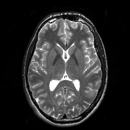 Here's a T2-weighted axial image. The third ventricles are seen in white. You can also see the caudate and putamen, separated by the anterior limb of the internal capsule.
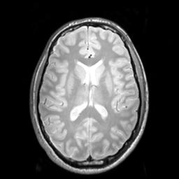 For comparison to the T2-weighted image, here's a proton density axial slice.
2 comments:
Those are such cool pictures. I want an mri scan too! :)
jane
Hey Jane
I know. If only the neonatal scans I'm working with were as good. Unfortunately, neonates have smaller brains with a much higher water content, and coupled with the fact that they have a tendency to move around in the scanner, images are of worse quality. We're working on it though!
Post a Comment