Showing posts with label Diffusion. Show all posts

Friday, December 16, 2005

Research update

A friend asked me to describe a little more about my project. So here goes! Let me know if you have any questions or would like to know more.

There is lots of evidence that the premature brain is particularly susceptible to injury that can result in long term problems including learning, behavioural and motor disabilities, and that it develops differently in the ex utero environment than it does in the womb. I'm looking at magnetic resonance brain images of preterm (i.e. premature) babies who were scanned at term equivalent age and comparing these to images from term born babies. I'm not using conventional MRI to compare them though, but with a technique called diffusion tensor imaging (DTI), where the image intensity is dependent on the diffusion properties of water within the tissue. Water molecules are restricted by microstructural boundaries including cell membranes and axonal tracts, which become more numerous as the brain develops with age. DTI therefore allows you to assess brain microstructure, and so may allow the visualisation of brain lesions in preterm infants before they are seen with conventional MRI. However, due to the way DTI data is acquired, it's susceptible to severe image distortion in a particular direction. I'm working on ways of computationally correcting this post-scan, so that the data is good enough to allow comparisons. I'm also trying to put all of the images into a common reference frame, so that the comparisons can be objective, unlike region of interest analyses.

It's probably easier to explain this last part if I show you a couple of pictures. In both images, which are taken from the same infant, a T2 MRI slice is on the left of the dotted line, and a corresponding B0 slice is on the right. I am trying to put them into the same reference frame. 'Before' shows alignment before I applied the registration, and 'After' shows alignment after registration. The yellow arrows reflect the tissue displacement that has taken place. It doesn't yet work as well all the time, but I'm hoping to improve that! Once I’ve got all the brains into a common reference frame I can begin comparing groups of preterm and term infants.

Anyway, I'd better hear off - it's our Christmas party. Should be fun!

All the best,
Moc

Before (structures misaligned)


After


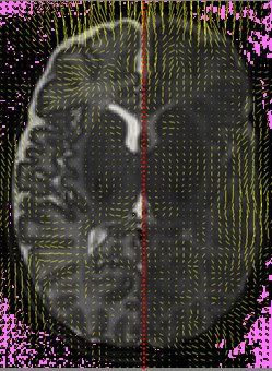
P.S. Please ignore all that pink noise - it just appeared when I tried to upload the images to blogger.

Thursday, September 01, 2005

The conference, day two

Yesterday afternoon there was a diffusion tensor imaging workshop as part of the conference, which was really useful for me, as that is the MRI modality I will be using throughout the PhD. It was valuable to know what’s going on in the world of MRI, and to speak to some of the people whose research papers I’m reading every day. I’m lucky in that respect because the postdoc we’re with is a bit of an imaging legend who has done important work and seems to know almost everyone. He was quite blunt during today’s parallel imaging session though, questioning the usefulness of the research done by a speaker and calling his signal-to-noise ratio “cr*p”!

What was your first conference/public speaking experience like?


Tuesday, August 30, 2005

PhD Proposal

My department of computing supervisor DR got back to me today and approved my PhD proposal. That means that I can start getting my data a month early, which is a huge bonus. Thanks!

Here’s my proposal in very truncated form. I've had to leave out all the references, study design, methods and technical sections. At least it doesn’t contain all the scary physics and equations of the full document though! I hope it makes it a bit clearer what I’ll be working on for the next three years. Please leave comments (click on the comments hyperlink at the bottom of the text) and feel free to ask any questions or request the whole document.

Yours,
Moc


1. Background
Over the past decade the incidence of low birth weight (infants weighing less than 2500g) has increased steadily, and now represents over 7% of all live births in England and Wales. Over the same period the incidence of very low birth weight (i.e. infants weighing less than 1500 grams) has increased at an even faster rate, now making up over 1% of all births. Although the outcomes for these infants vary widely across different neonatal intensive care units, recent improvements in perinatal care now mean that around 90% of these preterm infants will survive. The improvements in survival have been greatest in the most immature infants but have been accompanied by an increasing awareness of subsequent neurodevelopmental deficits. The immature developing brain is very vulnerable to injury, and many preterm infants suffer long-term morbidity that is more severe with prolonged exposure to the extrauterine environment. Impairments often continue into adolescence, with a high prevalence of behavioural problems documented, including psychiatric and attentional deficit disorders.

Magnetic resonance imaging (MRI) has been increasingly used within the field of neonatology over the past decade. Indeed, most of the recent insights into intrauterine and early extrauterine brain development have been achieved thanks to conventional (T1- and T2-weighted) magnetic resonance imaging techniques. MRI findings correlate with the well-recognised pathologies seen on ultrasound and, in addition, the high spatial resolution and excellent soft tissue contrast also allows the detection of more subtle abnormalities, including increased extracerebral space and diffuse excessive high signal intensity.

Diffusion tensor imaging (DTI) and diffusion weighted imaging (DWI) are magnetic resonance techniques that provide quantitative measures of water diffusion in tissue. By doing so they are able to show brain physiology and microstructure in vivo. The image contrast in DWI and DTI depends on the diffusion characteristics of water molecules, which are restricted by structural barriers including cell membranes and white matter tracts. Values of the apparent diffusion coefficient and fractional anisotropy can be determined from DTI and, by calculating the eigenvalues of the diffusion tensor, diffusion parallel and perpendicular to the white matter tracts can be measured. These are non-subjective measurements and provide information reflecting tissue microstructure. They can therefore be used to assess micro-structural abnormalities in the preterm brain.

Computer-assisted morphometric techniques, including voxel-based morphometry (VBM) and deformation based morphometry (DBM) use image registration and statistical analysis to quantify structural differences between groups To date, there have been no studies applying DBM techniques to diffusion scalar maps (apparent diffusion coefficient, fractional anisotropy and eigenvalue maps) in the preterm brain. However, data from animal and adult studies suggest that combining these two techniques (DBM and DTI) provides information regarding microstructural anomalies that may not be observed using traditional region of interest analysis of DTI data.

In this project I aim to use diffusion tensor imaging and deformation based morphometry to assess microstructural abnormalities in the preterm brain.

2. Hypotheses
i. Deformation based morphometry of diffusion tensor imaging data is able to detect abnormal white matter development in infants who are born prematurely.
ii. Abnormal white matter development is the result of extreme preterm birth and is not associated with other events.


3. Aims
i. To compare diffusion tensor scalar maps of the preterm brain at term equivalent age to those of infants born at term using DBM.
ii. To assess the effect of gestational age at birth on white matter development by acquiring DTI data at three time-points for each preterm infant: within 1 week of birth, at 30 weeks GA and at term equivalent age.
iii. To investigate the effect of other factors on white matter development including antenatal infection, postnatal infection, chronic lung disease, intrauterine growth retardation and gender.